A Comparison of Radionuclide Ɣ-ray Irradiators and Orthovoltage X-ray Irradiators
Many high-energy irradiator systems use gamma rays, which are generated through the decay of radioactive material. Though high-energy irradiators yield high energy photons of a single energy (monoenergetic), these systems have important safety concerns which have resulted in a U.S. government program called CIRP (Cesium Irradiator Replacement Project) that incentivizes the replacement of these systems with safer technology such as X-ray radiation. In Europe, the EU’s nuclear safety framework supports gamma source reduction, and some European countries have national programs with similar objectives.
Some disadvantages of gamma irradiation systems are listed below:
- The radioactive material present in gamma sources requires personnel with specialized training who have undergone thorough background checks to manage and uphold strict safety guidelines.
- Radioactive sources must be monitored and regulated by relevant nuclear regulatory institutions.
- Radioactive sources are always active. Nuclides decay constantly; there is no way to “turn them off”. A mechanical malfunction of the cabinet can result in a serious environmental radiation hazard.
- The amount of radiation from radioactive sources reduces over time. There comes a point where the radiation levels fall below certain usable thresholds, and the irradiator must be decommissioned.
- Decommissioning a radiation source is expensive and complicated, and the radioactive material persists for hundreds of years.
Conversely, orthovoltage irradiators generate x-rays through the acceleration of electrons that impinge on a metal surface. Advantages of these systems include:
- X-ray cabinet irradiators do not require specialized personnel to operate them. Researchers and students may operate them with minimal training.
- X-ray sources can be turned off when they are not being used, lessening the risk of accidental exposure, and virtually eliminating the risk of an environmental radiation hazard.
- Even though x-ray sources produce poly-energetic beams, a portion of the lower energy photons may be attenuated through the appropriate use of filters, making the photon spectrum more homogeneous.
- Decommissioning an x-ray source is easier and less expensive since there is no radioactive material to deal with.
- The dose rate is adjustable and remains constant at fixed voltage and current during the source’s effective life.
It is important to note that published data shows that the uniformity of dose deposited in soft tissue is independent of irradiation method, whether γ-ray, megavoltage, or x-ray sources are used [1].
Below are some additional factors to consider when switching from gamma to X-ray irradiation:
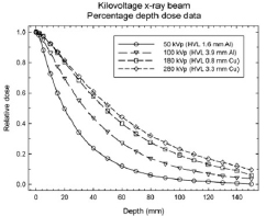
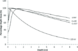
- Penetration depths as well as dose rates will be different between Cesium and X-Ray Irradiator (See Figures 1 [2] and 2 [3]).
- Studies have shown marked differences in the relative biological effectiveness (RBE) and discrete cellular responses to gamma vs X-ray radiation* [4]
- Lower energy (soft) x-rays increase the superficial dose to tissues (no skin-sparing effect). However, these energies can be filtered appropriately.
- A 320 kVp x-ray source has a dose rate that is 40% lower than a Cs137 source [1]. This reduced dose rate can be compensated for by increasing the irradiation time.
- An x-ray beam can be adjusted to yield any desired output (dose rate) within the operating range of the irradiator, by adjusting the voltage and current.
References
[1] Caravaca, J., Peter, R., Yang, J., Gunther, C., Serrano, J. A. C., Nostrand, C., ... & Seo, Y. (2022). Comparison and calibration of dose delivered by 137Cs and x-ray irradiators in mice. Physics in Medicine & Biology, 67(22), 225017.
[2] Hill, R., Healy, B., Holloway, L., Kuncic, Z., Thwaites, D., & Baldock, C. (2014). Advances in kilovoltage x-ray beam dosimetry. Physics in Medicine & Biology, 59(6), R183.
[3] Koontz, Bridget & Verhaegen, Frank & Ruysscher, Dirk. (2016). Tumour and normal tissue radiobiology in mouse models: How close are mice to mini-humans? The British journal of radiology. 90. 20160441. 10.1259/bjr.20160441.
[4] Shahmohammadi Beni M, Krstic D, Nikezic D, Yu KN. Realistic dosimetry for studies on biological responses to X-rays and γ-rays. J Radiat Res. 2017;58(5):729-736. doi:10.1093/jrr/rrx019
