When evaluating preclinical irradiators, there are a few considerations that can help you decide which X-ray irradiation system has the characteristics that will best suit your needs.
Some basic considerations such as features, footprint (benchtop or free-standing), price, reliability, and after-purchase support are all important factors. Another consideration you may not be aware of is beam quality.
What is Beam Quality?
Beam quality defines the penetration capability of an X-ray irradiator’s beam. It is described by two characteristics:
- Average beam energy
- Beam homogeneity
X-rays are created when high-speed electrons release energy in the form of photons. Every X-ray beam is made up of photons with a spectrum of energies. ‘Average beam energy’ is exactly what it sounds like; the average energy of all the photons in an X-ray beam. Different system configurations will achieve different average energies which are mainly dependent on two factors; peak energy of the beam and beam filtration.
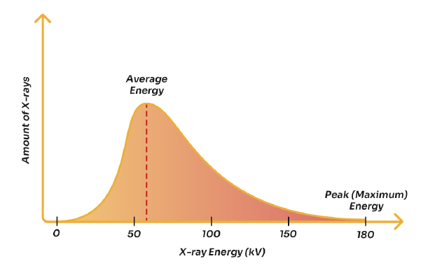
- Peak energy (kV) is the highest energy that a system can deliver. It is a function of the maximum potential energy a unit can generate. It’s important to note that an x-ray tube will generate x-rays of its maximum potential energy as well as all energies below that max. (see Figure 1).
- Beam filtration is a method for removing lower energy photons and retaining higher energy photons, thereby increasing the average energy of the beam.
These two factors can shift a beam’s energy spectrum to a higher average energy, which is one of the characteristics of a higher quality beam. (We’ll discuss beam homogeneity in the next article…)
Beam quality is not a function of the amount (or flow rate) of electrons, which is dictated by the current (mA). Increasing the mA will increase the quantity of electrons and photons, thereby increasing the dose, but it does not change the energy, or penetrating power, of the beam (i.e. the depth at which the dose is delivered). Dose delivery at depth is a function of beam quality and is mainly dependent on kV and filtration -- and not on mA or exposure time.
The Importance of Beam Quality
Beam quality is an extremely important consideration, especially when the radiation dose is being delivered into a biological system. When the target is a tumor inside an animal and the system configuration produces a low-quality beam, it will have a larger spectrum of energies that will include lower energies. Lower energy x-rays will not penetrate tissues deep enough to reach the tumor; but can instead cause burns to the skin, because such beams have very little to no penetration ability. A high-quality beam, on the other hand, will have a higher average energy due to the removal* of lower energy photons and a higher peak energy. This will allow for better depth of penetration, with less surface-level damage and more of the beam reaching the tumor.
“Higher” and “lower” here are relative terms. Generally, X-ray irradiation systems currently on the market can achieve peak energies ranging from 150 kV to 320 kV. (It may be of interest to note that X-ray systems specifically for imaging generally have peak energies that range from 20 kV to 90 kV, delivering a much lower radiation dose and requiring a much shorter exposure time for imaging as compared to therapeutic radiation dosing.) The needs of your study, lab, department, organization, or budget will help determine the system and appropriate kV for your situation.
In the next installment, Beam Homogeneity: A Contributor to Beam Quality, we'll dive deeper into that second factor of beam quality, the homogeneous beam. We’ll explore what factors contribute to it and why it matters....
*Lower energies are removed using filtration; a method we’ll discuss in later articles.
