Three Critical Considerations for Preclinical X-ray Imaging
When evaluating preclinical X-ray imaging systems, it can be helpful to understand the factors that make for a quality system. We've broken it down into three categories that we'll introduce here and explore further in the article links to the left.
High-Performance X-ray Components: Source kV and Detector Focus
First, the power and sensitivity of the instrument components (i.e., source kV and detector focus) are key. X-ray components must be capable of capturing and generating high-resolution images, otherwise no amount of image optimization can compensate when detailed information is lacking from the start.
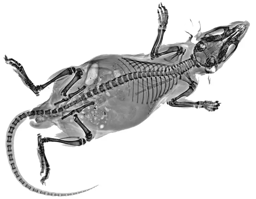
Applications for High-Resolution Visual Examination
Capturing fine details enables accurate visual examination and analysis of meaningful biology, including:
- Bone Research: Structure and density of trabecular bone, skeletal development, and fracture lines/longitudinal bone remodeling.
- Soft Tissue Characterization: Intervertebral disc space, visualizing fibrotic tumors, and more.
There should be no compromise on a system's ability to generate images where the level of clarity needed to corroborate findings from other imaging modalities is provided.
Intuitive Imaging Tools and Advanced DXA Software
Second, intuitive imaging tools and innovative software that complement imaging capability make for a complete imaging system, capable of generating more robust, quantitative datasets.
For example, next-generation DXA software that can simulate CCA (Carcass Composition Analysis) and NMR (Nuclear Magnetic Resonance) brings a multitude of advantages to the lab:
Automated Image Processing and Manual Optimization
Finally, effective pre- and post-image processing can ensure images are automatically optimized based on the characteristic density of the sample.
Empowering Users with Custom Imaging Control
Users may wish to further optimize an image based on their own specific imaging goals. A quality system offers a balance of:
- Automatic Optimization: For speed and consistency across samples.
- Manual Optimization: To empower users to obtain the specific visual information they need quickly and easily.
Conclusion: Hardware, Software, and Optimization
System performance, software intelligence, and optimization flexibility are the three pillars of effective X-ray imaging.
