A Better Beam
Made up of a spectrum of energies, an X-ray beam that has its spectrum shifted to higher average energy will have improved beam quality, a desirable trait for performing research irradiation. Recall from our earlier articles that the quality of the beam is described by two beam characteristics: how evenly the radiation dose is distributed (both its spatial uniformity across the area of a radiation field and its depth uniformity), and its average energy. Shifting a beam’s energy spectrum to a higher average energy can be achieved by taking advantage of two properties of an irradiation system:
- Kilovoltage (kV): A measure of the peak energy within the spectrum of energies in an X-ray beam. It follows that the average energy will be higher when using a system capable of achieving a higher peak energy than when using a comparable system that can achieve a lower peak energy (“higher” and “lower” here are relative terms because the energy level should still be appropriate for its intended use).
- Beam filtration: A method for removing lower energy photons and retaining higher energy photons, thereby reducing the overall energy spectrum and increasing the average energy of the beam.
Beam filtration is the key to removing lower energy photons and increasing the quality of the beam. This is true for all X-ray irradiator systems regardless of their peak kV.
What is an X-ray Filter?
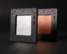
An X-ray filter is a material that is placed in the path of an X-ray beam and preferentially blocks low-energy photons from passing through while allowing higher-energy photons to pass. Filters are made from metals of different densities, such as aluminum, copper, or tin, and vary in thickness, which together will determine the specific filtering ability. All irradiation systems have inherent filtration, which is a filter that has been built into the system and cannot be removed. Selectable filtration, on the other hand, can be added or removed as needed, based on sample type and experimental needs. KUBTEC® irradiators have both inherent filtration and an array of selectable filters, both standard and optional.
In practice, using filters means that a beam will penetrate to a more uniform depth with less energy scattering at the surface of the sample being irradiated. When the material being irradiated is biological, this provides advantages that include reduced risk of skin burns and a more penetrative, predictable, and reproducible dose.
Comparing Two Systems

On nearly any X-ray irradiation system, adding filtration will increase beam quality to some degree. However, within the energy level range that is appropriate for the intended use, the ability of filtration to improve beam quality is limited by the peak energy of the system, and other factors as well.
Reducing the spectrum of energies of both systems by using filtration will shift both X-ray spectra toward higher potentials and yield higher average beam energies; factors of improved beam quality. However, with the appropriate filters applied, the beam quality achieved is greater if a higher peak energy is attainable. Therefore, the system capable of delivering up to 180 kV will provide a higher beam quality, which an irradiator delivering up to 150 kV will be unable to replicate.
There are other considerations that can further improve beam quality. To sharpen the distinction between the two systems in our comparison, the 180 kV system incorporates a novel design that enables it to generate a more homogeneous beam* such that, when selectable filtration and rotation are added, the system generates a uniquely high-quality beam that can meet the needs of cell or small animal research studies.
With the right understanding of why irradiation filtration matters, and what other factors can influence beam quality, researchers will be empowered to generate data that is more reproducible and more robust for better research outcomes.
In the next installment, Determining X-ray Beam Quality, we'll explore what a Half-Value Layer is and how it functions as a method to measure beam quality....
